Adrenal Anatomy
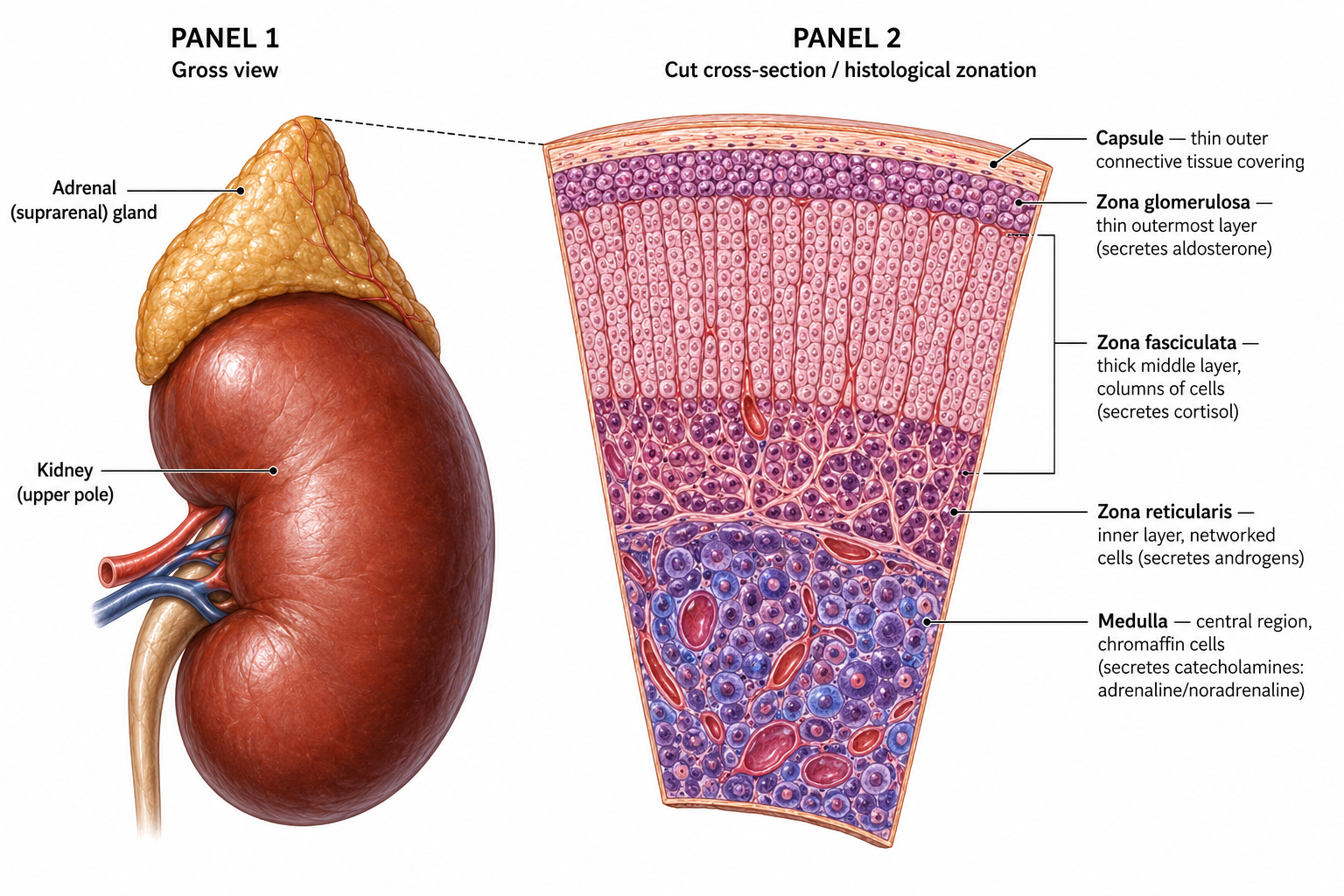
Each kidney is capped by an adrenal gland — a three-layered cortex (aldosterone, cortisol, sex steroids) over a catecholamine-secreting medulla — and the surgically load-bearing fact is the short right adrenal vein draining straight into the IVC.
The big picture
Each kidney is capped by an adrenal gland, both enclosed in Gerota's (perirenal) fascia; ~4–5 g each (right triangular, left crescentic). The cortex (85%) has three layers, each with a distinct endocrine job: zona glomerulosa → mineralocorticoids (aldosterone), zona fasciculata → glucocorticoids (cortisol), zona reticularis → sex steroids; the medulla secretes catecholamines.
Cortical zonation explains the hormones; the short right adrenal vein → IVC explains the surgical danger.
Mechanism pathway
Tap any step to see why it happens.
Adrenal gland — gross view and histological zonation
Board traps
Right adrenal vein → IVC (short); left adrenal vein → left renal vein. Same pattern for testicular veins.
Adrenal cortex layers: glomerulosa→mineralocorticoid, fasciculata→glucocorticoid, reticularis→sex steroid ("salt, sugar, sex").