Testicular Cancer
A solid testicular mass is cancer until proven otherwise — ultrasound, markers before orchiectomy, radical inguinal orchiectomy, then treat by seminoma-vs-NSGCT and stage.
The big picture
Think of a fixed sequence: feel a mass → ultrasound confirms it is intratesticular and solid → tumour markers are drawn before surgery → radical inguinal orchiectomy gives diagnosis and local control → histology (seminoma vs non-seminoma) and stage drive the rest.
Solid intratesticular mass = radical inguinal orchiectomy; never trans-scrotal, and always markers before surgery. AFP elevation means it is NOT a pure seminoma.
Mechanism pathway
Tap any step to see why it happens.
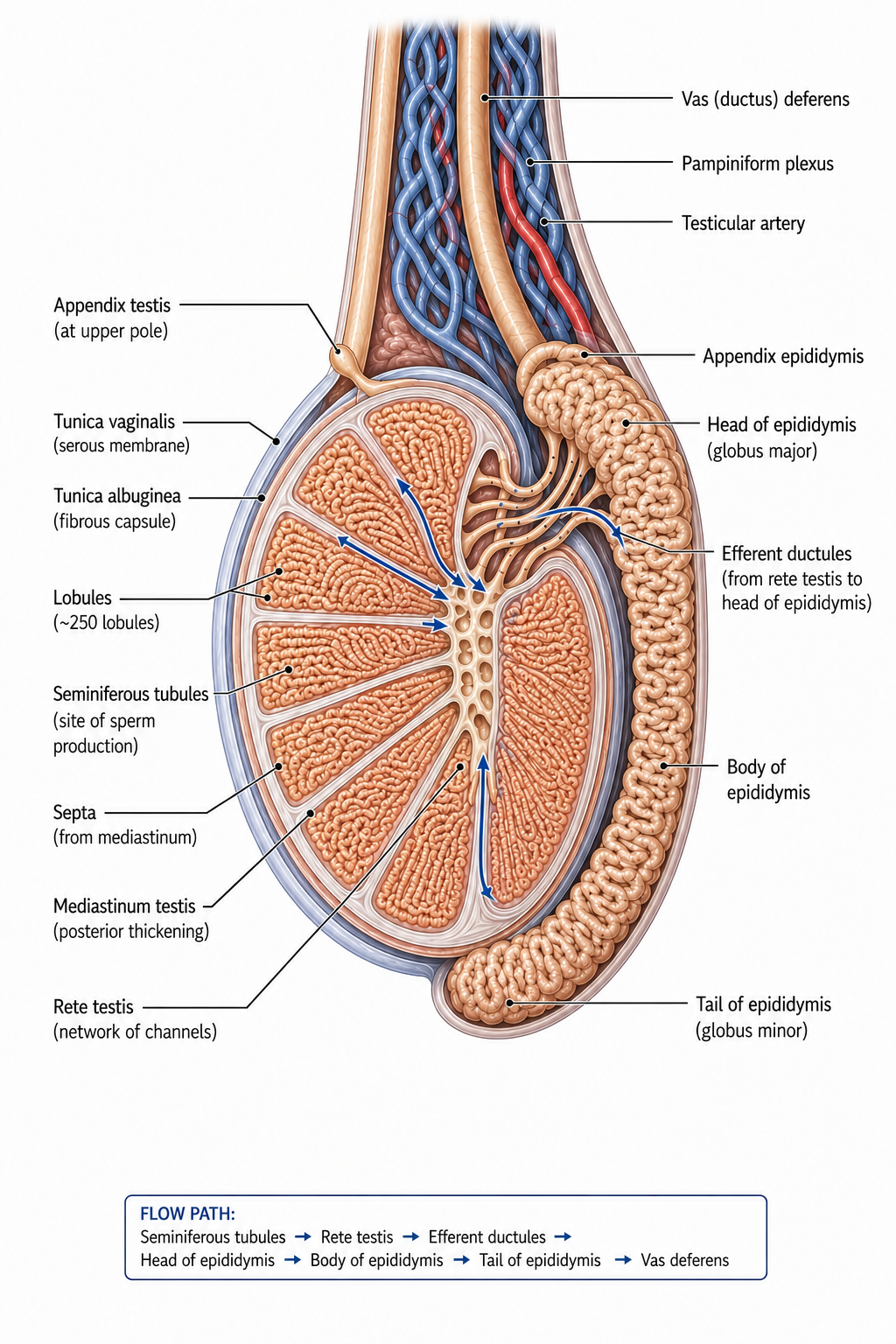
The testis & epididymis
Interactive — testis cancer post-orchiectomy management
Diagnostic algorithm
Each step answers one question. Tap to expand.
Classification & subtypes
Seminoma
Pure germ cell tumour, radiosensitive and chemosensitive.
- Why it matters:
- AFP is NEVER raised in pure seminoma; mild hCG can occur.
- Management:
- Stage I: surveillance or single-agent carboplatin. Advanced: platinum chemotherapy.
- Memory hook:
- Seminoma + raised AFP is impossible — treat as NSGCT.
- Board trap:
- A 'seminoma' on histology with high AFP is a non-seminoma.
Non-seminomatous GCT (NSGCT)
Embryonal, yolk sac, choriocarcinoma, teratoma — alone or mixed.
- Why it matters:
- AFP and/or hCG often raised; more aggressive, chemo/RPLND-based.
- Management:
- Stage by markers/nodes; BEP chemotherapy and/or retroperitoneal lymph node dissection.
- Memory hook:
- AFP up = NSGCT; choriocarcinoma drives very high hCG.
- Board trap:
- Teratoma is chemo-resistant — residual masses may need RPLND.
Marker logic (AFP/hCG/LDH)
Markers classify, stage (S category) and monitor response.
- Why it matters:
- Rising markers after orchiectomy mean residual/metastatic disease.
- Management:
- Draw before surgery, repeat after, and trend during treatment.
- Memory hook:
- AFP = yolk sac/embryonal; hCG = choriocarcinoma; LDH = burden.
- Board trap:
- Failure to draw pre-op markers loses staging information.
Treatment ladder
Diagnosis and local control by inguinal orchiectomy first; then type-, stage- and marker-directed therapy — with cure as a realistic goal even when metastatic.
Procedure chooser
- Radical inguinal orchiectomy (diagnostic + therapeutic)
- Resection of residual masses after chemotherapy
Follow-up
- Tumour markers (AFP, hCG, LDH) trend
- Cross-sectional imaging on a stage-based schedule
- Late effects of chemotherapy/radiation
- Intensive in the first 2–3 years (highest relapse risk), then tapering
- Marker check at each visit
- Markers normalise and stay normal; no new disease on imaging
- Rising markers or new nodal/visceral disease (relapse)
- Per surveillance protocol and for rising markers/symptoms
- Second malignancy and cardiovascular risk after chemo/radiation
- Hypogonadism/fertility
- Relapse in teratoma elements
Memory hooks
US → markers → inguinal orchiectomy → stage.
AFP = NSGCT (yolk sac/embryonal).
Pure seminoma: AFP normal.
Teratoma resists chemo — cut it out.
Never go trans-scrotal.
Board traps
Histology says seminoma but AFP is high — treat as non-seminoma.
Residual mass after chemo in NSGCT — teratoma; resect it.
Trans-scrotal approach taken — altered drainage, worse staging.
Gynaecomastia + testis mass — hCG-secreting tumour.
Markers not drawn before orchiectomy — staging compromised.
Clinical cases
A 27-year-old man has a painless firm right testicular mass. Ultrasound confirms a solid intratesticular lesion. AFP is elevated; hCG mildly raised. Histology after orchiectomy is reported as 'seminoma'.
How should this be classified and managed?