Bladder Anatomy
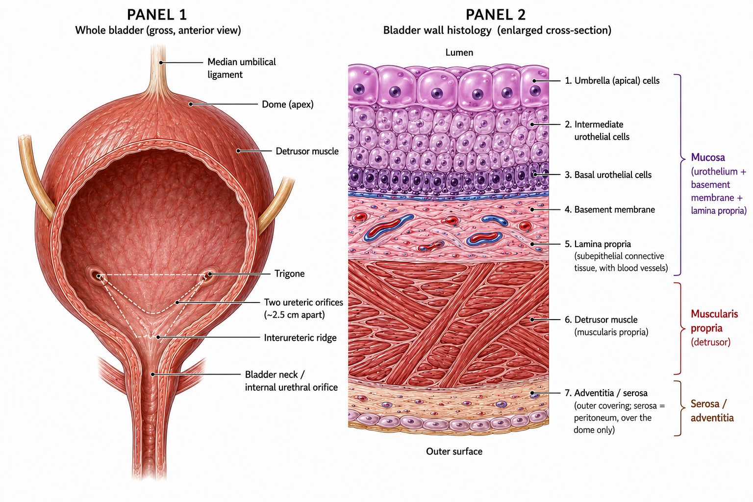
A layered muscular reservoir — urothelium → lamina propria → detrusor → adventitia/serosa — whose wall layers are the anatomical basis of bladder-cancer staging, with the trigone marking the antireflux base.
The big picture
Hollow muscular reservoir, ~400–500 mL. The trigone is the smooth triangle between the two ureteric orifices and the bladder neck; ureters enter obliquely (a flap-valve preventing reflux). The urachus runs from dome to umbilicus (→ median umbilical ligament). The bladder is lined by transitional epithelium (urothelium) — the same lining from calyx to bladder.
Wall layers = staging: Ta (mucosa) → T1 (lamina propria, non-muscle-invasive) → T2 (detrusor, muscle-invasive).
Mechanism pathway
Tap any step to see why it happens.
Bladder — whole organ & wall histology
Board traps
Bladder wall (inside-out): urothelium → lamina propria → detrusor (3 muscle layers) → adventitia/serosa.
Staging maps to layers: Ta = mucosa, T1 = lamina propria (non-muscle-invasive), T2 = detrusor (muscle-invasive).
Trigone = between the two ureteric orifices (~2.5 cm apart) and the bladder neck; distinct embryologic origin.